Home » 3D Imaging

At Oxford OMS Centre, we understand that accurately diagnosing each patient’s individual condition is critical to successful surgical outcomes. While most conditions can be fully assessed with a history, clinical examination and two-dimensional (2D) imaging, certain conditions require three-dimensional (3D) imaging. In these more complex cases, 3D imaging will allow a thorough evaluation of the teeth, bone, and/or vital structures in the area of interest.
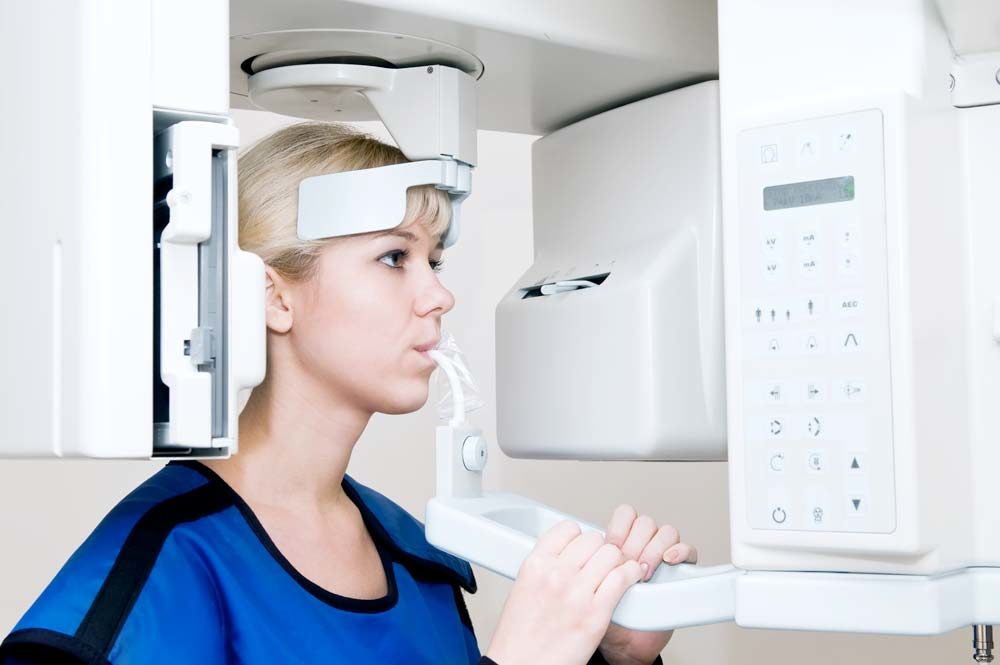
Our 3D imaging system (Axeos | Dentsply Sirona) utilizes cone beam technology, like a mini-CT scanner, to provide high-resolution images of the dental/facial tissues. These images are acquired at a much-reduced dose of radiation compared to medical grade CT scanners without compromising diagnostic quality. Once a scan has been taken, it is sent to our maxillofacial radiologist for interpretation, and we are sent a detailed report to add to each patient’s medical record.
Examples of clinical situations which may require 3D scanning include: dental implant planning, oral/maxillofacial pathology, airway space evaluation, pre-orthodontic treatment, pre-corrective jaw surgery, evaluation of impacted teeth, maxillary sinus augmentation, and other bone grafting procedures.
© Copyright Dr. Matthew D. Morrison, Oxford Oral & Maxillofacial Surgery Centre | Dental Website Design by Platinum Design